SAMPLES REQUIRE IDENTIFICATION

SAMPLES REQUIRE IDENTIFICATION

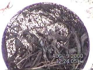
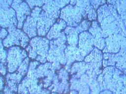
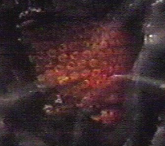
Three separate samples that were collected in August 2000 are pictured and discussed in this paper. The first sample analyzed was sent in from Missouri and is a dark brown/black powdery substance, and is possibly a fungus. A microscope shows the substance to be made up of uniform spherical structures about 7 microns in diameter. The second substance discussed is a fibrous material that was found in a previous rainwater sample collected on June 26-27, 2000 in Santa Fe, New Mexico. When dried, these fibers appeared dendritic in nature, and under a microscope, they measured roughly 1 micron in diameter. The third substance is a material that appeared to have originated from the ground in Albuquerque, New Mexico, and is not believed to be related with aerial activity. This material, described as having a foam-like nature and a texture like ‘chocolate mousse’, is believed to be fungus related, though such a growth is not known to thrive in desert conditions in New Mexico. Input is welcome in determining what these substances are. Those with mycology or microbiology backgrounds are encouraged to respond to the Carnicom Institute.
ADDITIONAL BIOLOGICAL COMPONENTS IDENTIFIED

ADDITIONAL BIOLOGICAL COMPONENTS IDENTIFIED

Microscopy stills are attached in this paper from fiber samples that had been previously evaluated in an earlier Carnicom paper named BIOLOGICAL COMPONENTS IDENTIFIED that was published on May 11, 2000. Though a portion of this same fiber sample was sent to EPA Administrator Carol M. Browner six months prior to this paper, to date, Ms. Browner refuses to identify the material in this sample.
INQUIRY : RAINWATER ANALYSIS

INQUIRY : RAINWATER ANALYSIS

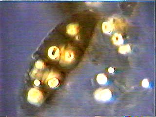
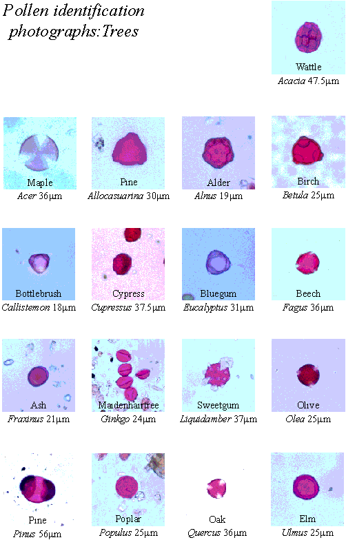
Two separate rainwater samples collected during the first substantial rains in Santa Fe, New Mexico on June 26th and 27th are analyzed under a microscope in this discussion. Both samples appear to show non-moving, double celled structures measuring 40-50 microns in diameter and showing up as separate structures and in clusters. After posting on the www.carnicom.com message board eliciting input from those with microbiology or microscopy experience in identifying these structures and receiving no responses, these structures were assumed to be pine pollen and discussion was discontinued. However, after receiving an anonymous response on the message board stating that pine pollen is used in clandestine genetic engineering experiments, this discussion was reopened. Anyone with microscopy and/or microbiology backgrounds who can help identify these structures, or give other relevant information regarding these structures, are invited to contact the Carnicom Institute with any and all data points to help in this regard.
BIOLOGICAL COMPONENTS IDENTIFIED

BIOLOGICAL COMPONENTS IDENTIFIED

Further testing of ground fiber samples previously collected and analyzed (see papers titled AEROSOL GROUND SAMPLES) revealed biological components in the fibers - numerous red blood cells, white blood cells, and unidentified cell types have been found in the ground samples. The red blood cells, readily visible after being subjected to immersion oil, appear to possibly be of a freeze dried or desiccated nature. Numerous pictures from the microscope video show these biological as well as unidentified components. The surfaces of the cells appear to be modified in some way, but electron microscopy will likely be required to establish further detail.
ULTRAVIOLET LIGHT INVESTIGATIONS

ULTRAVIOLET LIGHT INVESTIGATIONS

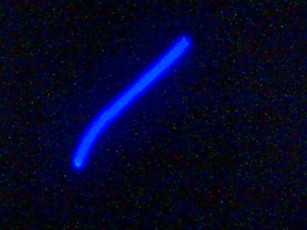
A ground fiber sample received previously in November 1999 is observed and pictured in this paper under a microscope and black light, and is compared to other known and unknown synthetic fibers. A notable thing from this observation is that the ground fibers show a low level of fluorescence, whereas the synthetics compared to show a high level of fluorescence. The ground fiber samples also showed sub-micron diameter, and high levels of adhesiveness and elasticity. Additional data and feedback of the current investigations underway with UV light is most welcome.
THIRD GROUND SAMPLE RECEIVED

THIRD GROUND SAMPLE RECEIVED

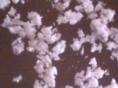
Pictures on this page show unusual samples from Sacramento, California that a witness sent in to the Carnicom Institute. These samples, which are white and of granular or powder form, are not at all similar to the fibrous microscopic samples sent in and discussed earlier. The significance of this ground sample, as well as the identification of it, remains unknown at this time.
AEROSOL GROUND SAMPLE: MICROSCOPIC VIEWS

AEROSOL GROUND SAMPLE: MICROSCOPIC VIEWS

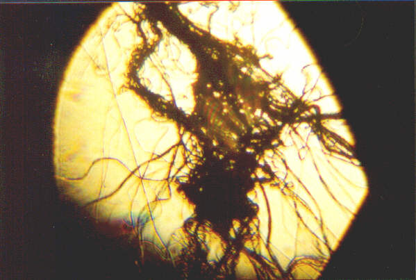
Microscopic views are presented of two filaments taken from a ground fiber sample after aerial spraying in eastern Oregon on November 2nd and November 4th, 1999. Observation and analysis indicate that the samples appear to be a polymer of some type, being both extremely elastic and adhesive, raising the possibility that this material may act as a carrier mechanism. The materials are white, and look like spider webs. The materials, under magnification, show individual strands that are wave-like in nature, and tend to coalesce and congeal easily. Ill health effects have been reported in association with the handling of this material. This material is reported to dissipate within a few hours of falling on the ground, and in being exposed to the weather. The ground fiber sample images are compared to and found not to be spider webs, and to be fully synthetic. Common health effects associated with this spraying include severe respiratory problems, burning eyes, feeling tired, and some people coughing up blood.
AEROSOL GROUND SAMPLES : MICROSCOPIC FIBERS REVEALED

AEROSOL GROUND SAMPLES : MICROSCOPIC FIBERS REVEALED

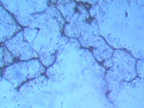
Eyewitness accounts of finding unusual fiber materials on the ground have been accumulated over the past year and more in direct connection with unusual aircraft activity. As might be expected, there are repeated, frequent and widespread accounts of respiratory distress and allergic reactions reported in association with such aircraft activity. This paper continues previous research on and presents microscopic views of suspected chemtrail ground samples collected in November and December 1999. Two identical ground fiber samples (one from Sacramento, CA, and one from eastern Oregon) were received, analyzed and compared to synthetic and natural fibers, such as human hair, wool, silk, spider webs, cotton and more. Though found in locations hundreds of miles apart, these two ground fibers exhibited identical characteristics in all respects down to the microscopic level. Based on tests outlined in this paper, it has been demonstrated that the ground sample fibers cannot be identified as any known or common natural or synthetic fiber.