DNA CULTURE RESULTS

DNA CULTURE RESULTS

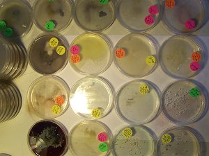
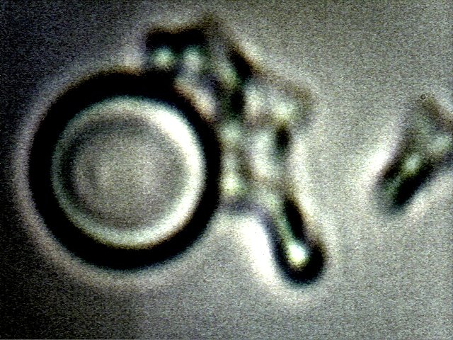
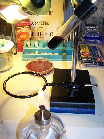
A method of extracting DNA samples from living forms has been established. The protocol being followed is that from the Genetic Services Learning Center at the University of Utah1. The methods have been applied successfully to human and fruit samples. Equipment to examine the internal structure of the DNA samples is not available at this time; visual microscopy techniques at relatively high magnification (~10,000x) are available.
MORGELLONS : AN ENVIRONMENTAL SOURCE

MORGELLONS : AN ENVIRONMENTAL SOURCE

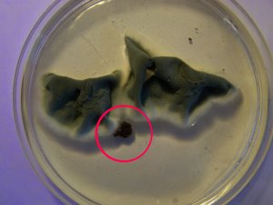
A continuing discussion of the characteristics of filament samples discovered by Clifford Carnicom and others is presented here. It is reiterated that an environmental source, at least in part, for specific biological organisms that are under scrutiny in association with the so-called "Morgellons" condition, has been identified. This source is the unusual airborne filament sample that was sent in June of 2000 to the Administrator of the United States Environmental Protection Agency (EPA) for identification on behalf of the public welfare. This particular and same sample that was sent to the EPA has been successfully cultured and reproduced, and the culture growth exhibits the identical biological organisms, structure and chemistry of certain biological filaments that are under extensive study in association with the Morgellons condition. The different cultured structures discussed above are described and pictured within this work as well. They are: 1) An encasing filament structure (containing an internal network of sub-micron filaments) 2) A chlamydia-like organism (Chlamydia pneumonia) 3) A pleomorphic form (Mycoplasma-like) 4) An erythrocytic form (red blood cell).
MORGELLONS : A STATUS REPORT

MORGELLONS : A STATUS REPORT

A partial summary of the research accumulated through this site on the so-called "Morgellons" issue is presented in this work. These summaries include fascinating, if not disturbing, details of the many years of work that Clifford Carnicom has put into his research that include the fact that: all individuals who participated in this research exhibit internal biological filaments and blood anomalies; the cultures produced from the internal biological filaments (dental samples of those participating in this research) have been shown to produce an erythrocytic (blood) form; and that the production of erythrocytic forms within direct biological filament samples and by culture is completely outside the known boundaries of conventional science and biology, and that it is repeatedly evident that these same erythrocytic forms can withstand (and even flourish in) extremely adverse environmental, chemical and thermal conditions. Many more details are provided in this long list of things known about Carnicom's work in this area.
ARTIFICIAL BLOOD (?)

ARTIFICIAL BLOOD (?)

Strong evidence now exists that an artificial or modified blood form is a dominant internal component, if not the dominant component, of dental filament samples that are commonly associated with the Morgellons condition. The sample set of this report is relatively small and it must be extended.  There is a remarkable consistency in the detailed observations and reports that have been made over a period of several years. This paper reaffirms the position of Clifford Carnicom that blood conditions and/or alterations appear to be at the crux of this situation. It is quite clear what type of work must be done to address the gravity of this situation, but additional resources must become available for this to take place. The current work now introduces the very real prospect or consideration that an artificial, or deliberately modified, process of the blood may have been introduced into the human condition. Elevated levels of research, aggressive involvement and appropriate resources must be dedicated and allocated to initiate progress on the many serious issues that have been disclosed.
MORGELLONS STATEMENT

MORGELLONS STATEMENT

This paper reiterates that there needs to be the clarification of the term 'Morgellons' to sway from originally being perceived as manifesting primarily an anomalous skin condition. Though there is a small portion of the population classified as having the Morgellon's condition, who show skin lesions that resist healing and have the presence of filaments that emanate from these sores, it is not the skin condition that defines the actual pathology. More recent research strongly indicates the underlying symptoms are much deeper and more broadly distributed than has been realized, and that blood borne vectors may be a common denominator amongst affected individuals. The presence of skin anomalies as the primary criterion for determining the existence of the condition appears to be especially deficient. Any reference to supposed "delusional parasitosis" in light of the physical examinations and documentation available appears to be a gross miscarriage and misdirection of effort to help these people, and needs to stop.
BLOOD ISSUES INTENSIFY

BLOOD ISSUES INTENSIFY

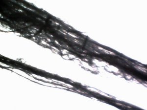
This paper discusses and presents applications of three independent methods established that appear to confirm the presence of developing modified erythrocytes (red blood cells) within cultured dental samples. These samples exhibit the characteristics of the Morgellons condition as previously researched and identified. All individuals tested thus far have produced the dental filamentous materials, regardless of whether visible skin anomalies are present or not. The erythrocytic detection methods are: 1.  Direct observation under the microscope at relatively high magnification (8000x - 10000x) using developed microscopy techniques. 2.  The use of the Kastle-Meyer presumptive test (visual and microscopic, sensitive test) for blood, a method commonly used in forensics for blood identification.   3.  The HEMASTIX (TMP) presumptive forensic test (very high sensitivity) commonly used for blood identification.
AEROSOLS & MORGELLONS: A Systems Perspective

AEROSOLS & MORGELLONS: A Systems Perspective

This page shows a systems perspective flow chart with the tie-ins of the aerosol programs and the Morgellon's symptoms and pathogens. Included in the chart's flows are headers such as Airborne Environmental Factors (Particulate, Filament and Biological Forms), Biological Findings (Morgellon's Condition - Blood, Skin, Hair and Dental), Culture Work and Aerosol Operations (Military, Environmental, Electromagnetic, etc.), and how they are related to each other in an easy to follow format.
MORGELLONS: GROWTH CAPTURED

MORGELLONS: GROWTH CAPTURED

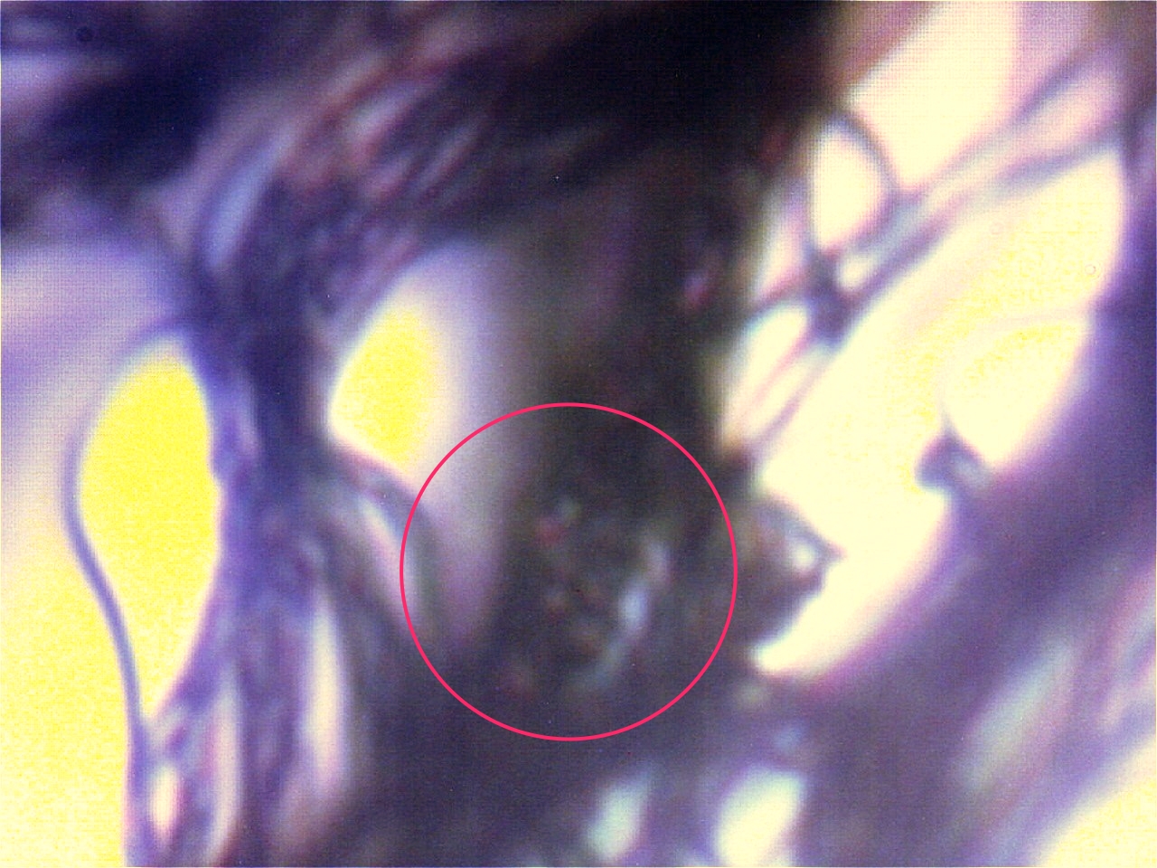
A time lapse video under the microscope has been developed and presented on this page which demonstrates the cultured growth pattern and behavior of a primary pathogenic form that is in direct association with the so-called "Morgellons" condition. The time lapse video covers a period of approximately six hours and compresses the time into approximately one minute with 30 frames. From the discovery shown here, it would appear that the encasing filament serves to provide feeder or extension filaments which serve to extend the growth of the pathogen. The estimated growth rate of the extension filaments on this particular culture is on the order of 50 microns per hour, or roughly the width of a thin human hair per hour. This report continues to add valuable knowledge on the morphology, characteristics and behavior of at least some of the pathogenic forms that are strongly associated with the so-called "Morgellons" condition.
CULTURE WORK IS CONFIRMED

CULTURE WORK IS CONFIRMED

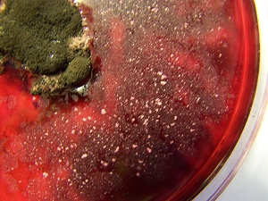
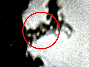
A pathogenic form that appears to be directly associated with the so-called "Morgellon's condition” has now been successfully, repeatedly and positively cultured from numerous independent dental filament samples over a protracted period of time. Confirmation of this result had been postponed until it became clear that the findings could be duplicated; this is now the case. High magnification microscopic images are provided to the reader for further understanding of what is being presented and the implications of this work. This work is important in that it provides a basis for the controlled study, observation, examination and modification of a primary pathogenic form that appears to underlie the existence of the so-called "Morgellon's" condition. It is reiterated that the general population appears to be subject to the existence of the pathogen, regardless of whether certain skin "anomalies" are present or not. As it is unlikely that Carnicom will have the time or resources to conduct the studies that are called for going forward in this research, it needs to be known that proper resources are a serious issue at this point. The public must now share in the responsibility for the progress (or the lack of it) that is dictated by this report.
CULTURE BREAKTHROUGH(?)

CULTURE BREAKTHROUGH(?)

Work has been conducted over the past one to two months that appears to be important and it may have significant impact on current research into pathogens being found in samples discussed in the most recent Carnicom papers.  It appears as though a primary pathogenic form under evaluation that is associated with the so-called "Morgellon’s" condition may have been successfully cultured.  If this proves to be the case, it offers the potential to begin very serious research on the methods to control, inhibit, reduce or eliminate the pathogenic forms within the human body. Unknown pathogens are difficult to identify, treat and remove if they exist only within the body; there is tremendous benefit if such pathogens can be grown or developed in a culture medium under controlled conditions.  This report may offer a pathway to that process. Further research at this time continues to show identical pathogens in samples that are viewed under an enhanced microscope setup, that without such enhancement, these images and what they reveal would not be possible. This would not have allowed the progress in this area of study by Carnicom that will possibly have tremendous positive implications for all of humanity.
Instructions for Boosting Your Microscope’s Power to Examine Your Own Samples

Instructions for Boosting Your Microscope’s Power to Examine Your Own Samples

Presented in this work are instructions for building an affordable yet fairly powerful microscope imager using a microscope and a typical webcam used with computers. This setup will allow anyone to get acceptable images of samples that have been discussed in the most recent Carnicom papers regarding the pathogens being found in people blood, gums, etc. A concerned citizen wrote to Carnicom in this paper, and describes using a Logitech Quick Cam, an empty film canister, and tape to enhance the magnification of a standard microscope by the use of a webcam's CCD sensor. It is noted that there is ample concern to have as many people reproduce Carnicom's results by the use of such a setup as is described here.